

 https://doi.org/10.4081/ahr.2026.207
The retinal age gap-particulate hypothesis: environmental exposure as a latent contributor to deep learning-based retinal age prediction
All claims expressed in this article are solely those of the authors and do not necessarily represent those of their affiliated organizations, or those of the publisher, the editors and the reviewers. Any product that may be evaluated in this article or claim that may be made by its manufacturer is not guaranteed or endorsed by the publisher.
Accepted: 8 June 2026
Published: 30 June 2026
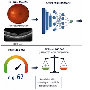
Deep learning models applied to retinal fundus photographs and optical coherence tomography (OCT) can predict chronological age with remarkable accuracy. The discrepancy between predicted and chronological age—the "retinal age gap”—is associated with mortality and multiple systemic diseases. However, the biological and physical correlates underlying these predictions remain incompletely understood. We propose that cumulative environmental particulate exposure—including microplastics, nanoplastics, and other airborne or ingested particulates—may contribute as a previously unrecognized component of the retinal age signal captured by deep learning models. Specifically, we hypothesize that particulate-associated retinal features, potentially manifesting as hyperreflective foci on OCT or tiny discrete foci on fundus imaging, may constitute one of several feature classes utilized by these models. We outline seven falsifiable predictions spanning epidemiology, imaging analysis, experimental manipulation, and post-mortem validation. Importantly, this framework does not posit that particulate burden is the primary determinant of retinal age, but rather that it may act as a latent, cumulative exposure signal embedded within a multifactorial aging phenotype. If supported, this hypothesis would expand the interpretability of retinal age models and position retinal imaging as a potential tool for studying long-term environmental exposure. This work is intended as a testable framework to guide empirical investigation rather than a definitive explanatory model.
Downloads
1. Poplin R, Varadarajan AV, Blumer K, et al. Prediction of cardiovascular risk factors from retinal fundus photographs via deep learning. Nat Biomed Eng 2018;2:158-64. DOI: https://doi.org/10.1038/s41551-018-0195-0
2. Zhu Z, Chen R, Yu Z, et al. Retinal age gap and the risk of age-related diseases. Invest Ophthalmol Vis Sci 2024;65:3769.
3. Wang Y, Liu T, Williamson D, et al. Age prediction from retinal fundus images and segmented vessel images using deep learning. Invest Ophthalmol Vis Sci 2023;64:1105.
4. Nielsen C, Souza R, Wilms M, Forkert ND. Foundation model-driven distributed learning for enhanced retinal age prediction. J Am Med Inform Assoc 2024;31:2550-9. DOI: https://doi.org/10.1093/jamia/ocae220
5. Yu Z, Chen R, Gui P, et al. A cross population study of retinal aging biomarkers with longitudinal pre-training and label distribution learning. NPJ Digit Med 2025;8:344. DOI: https://doi.org/10.1038/s41746-025-01751-7
6. Zhu Z, Shi D, Guankai P, et al. Retinal age gap as a predictive biomarker for mortality risk. Br J Ophthalmol 2023;107:547-54. DOI: https://doi.org/10.1136/bjophthalmol-2021-319807
7. Grimbly MJ, Koopowitz SM, Chen R, et al. Estimating biological age from retinal imaging: A scoping review. BMJ Open Ophthalmol 2024;9:e001794. DOI: https://doi.org/10.1136/bmjophth-2024-001794
8. Wu G, Zhang X, Borchert GA, et al. Association of retinal age gap with chronic kidney disease and subsequent cardiovascular disease sequelae: A cross-sectional and longitudinal study from the UK Biobank. Clin Kidney J 2024;17:sfae088. DOI: https://doi.org/10.1093/ckj/sfae088
9. Burlina PM, Joshi N, Pekala M, et al. Automated grading of age-related macular degeneration from color fundus images using deep convolutional neural networks. JAMA Ophthalmol 2017;136:1170-6. DOI: https://doi.org/10.1001/jamaophthalmol.2017.3782
10. Yee MS, Hii LW, Looi CK, et al. Impact of microplastics and nanoplastics on human health. Nanomaterials 2021;11:496. DOI: https://doi.org/10.3390/nano11020496
11. Leonard VL, Liddle CR, Atherall CA, et al. Microplastics in human blood: Polymer types, concentrations and characterisation using μFTIR. Environ Int 2024;188:108751. DOI: https://doi.org/10.1016/j.envint.2024.108751
12. Ragusa A, Svelato A, Santacroce C, et al. Plasticenta: First evidence of microplastics in human placenta. Environ Int 2021;146:106274. DOI: https://doi.org/10.1016/j.envint.2020.106274
13. Saha SC, Saha G. Effect of microplastics deposition on human lung airways: A review with computational benefits and challenges. Heliyon 2024;10:e24355. DOI: https://doi.org/10.1016/j.heliyon.2024.e24355
14. Wang YF, Wang XY, Chen BJ, et al. Impact of microplastics on the human digestive system: From basic to clinical. World J Gastroenterol 2025;31:100470. DOI: https://doi.org/10.3748/wjg.v31.i4.100470
15. Zhong Y, Yang Y, Zhang L, et al. Revealing new insights: Two-center evidence of microplastics in human vitreous humor and their implications for ocular health. Sci Total Environ 2024;921:171109. DOI: https://doi.org/10.1016/j.scitotenv.2024.171109
16. Zhang M, Liu S, Wang Y, et al. Detection and characterization of multiple microplastics in the human retina. Environ Sci Technol Lett 2025;12:1327-33. DOI: https://doi.org/10.1021/acs.estlett.5c00903
17. Zheng J, He L, Shi Q, et al. In vivo and in vitro assessment of the retinal toxicity of polystyrene nanoplastics. Environ Int 2025;198:109420. DOI: https://doi.org/10.1016/j.envint.2025.109420
18. Kadac-Czapska K, Ośko J, Knez E, Grembecka M. Microplastics and oxidative stress—Current problems and prospects. Antioxidants 2024;13:579. DOI: https://doi.org/10.3390/antiox13050579
19. Wang L, Pei W, Li J, et al. Microplastics induced apoptosis in macrophages by promoting ROS generation and altering metabolic profiles. Ecotoxicol Environ Saf 2024;271:115970. DOI: https://doi.org/10.1016/j.ecoenv.2024.115970
20. Silva MG, Oliveira MM, Peixoto F. The impact of micro-nanoplastics on mitochondria in the context of diet and diet-related diseases. Stresses 2025;5:12. DOI: https://doi.org/10.3390/stresses5010012
21. Tan AK. A Bayesian approach to investigating presumed retinal micro(nano)plastics. BioSystems 2025;253:105502. DOI: https://doi.org/10.1016/j.biosystems.2025.105502
22. Tan AK. A multi-modal artificial intelligence framework for identifying persistent exogenous-like hyperreflective foci in retinal optical coherence tomography. SSRN 2026. Available from: https://ssrn.com/abstract=5997994
23. Tan AK. Retinal hyperreflective foci as imaging biomarkers of environmental neurovascular stress: A scoping review of methodological approaches in clinically healthy eyes. J Clin Ophthalmol Res 2026;14:184-90. DOI: https://doi.org/10.4103/jcor.jcor_363_25
24. Manhas S, Anjali A, Malviya T, et al. Ubiquitous presence of microplastics with implication on soil health and plant development. Environ Monit Assess 2025;197:1043. DOI: https://doi.org/10.1007/s10661-025-14360-4
25. Ziani K, Ioniță-Mîndrican CB, Mititelu M, et al. Microplastics: A real global threat for environment and food safety: A state of the art review. Nutrients 2023;15:617. DOI: https://doi.org/10.3390/nu15030617
26. Aghabeigi Alooghareh MM, Sheikhey MM, Sahafi A, et al. Deep learning for comprehensive analysis of retinal fundus images: Detection of systemic and ocular conditions. Bioeng 2025;12:840. DOI: https://doi.org/10.3390/bioengineering12080840
27. Shahgir HS, Zaman TA, Sayeed KS, et al. Leveraging complementary attention maps in vision transformers for OCT image analysis. Proc. IEEE Int. Conf. on Image Processing (ICIP), Anchorage; pp. 1378-83. DOI: https://doi.org/10.1109/ICIP55913.2025.11084435
28. Zang B, Ding L, Feng Z, et al. CNN-LRP: Understanding convolutional neural networks performance for target recognition in SAR images. Sensors (Basel) 2021;21:4536. DOI: https://doi.org/10.3390/s21134536
29. Tan AK. Bayesian hypothesis generation: A probabilistic framework for evaluating novel hypotheses before data collection. Med Hypotheses 2026;206:111841. DOI: https://doi.org/10.1016/j.mehy.2025.111841
CRediT authorship contribution
Tan Aik Kah, conceptualization, methodology, software, validation, data curation, visualization, investigation, supervision, writing – original draft preparation, writing – reviewing and editing.
Data Availability Statement
not applicable
How to Cite

This work is licensed under a Creative Commons Attribution-NonCommercial 4.0 International License.
PAGEPress has chosen to apply the Creative Commons Attribution NonCommercial 4.0 International License (CC BY-NC 4.0) to all manuscripts to be published.